


By Dan Ross
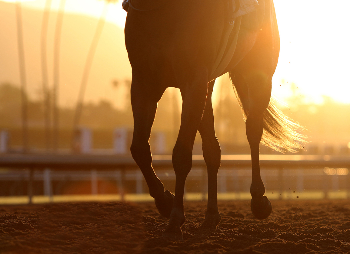
Seeking a full stop to the spate of high-profile fatalities in the race that stops Australia, Racing Victoria this year tightened the veterinary screws. The practical rollout of these efforts can hardly be described as an unadulterated success, however.
One of these new measures was a precautionary CT scan of all runners in the days leading up to the G1 Melbourne Cup–a target that hit the skids when Racing Victoria's new $1.27-million CT unit suffered a malfunction with the Cup field only half scanned, leaving the rest to be X-rayed (with a machine that was also temporarily incapacitated).
But other, less-mechanical incidents highlight some of the more nuanced problems that come with using sophisticated–and still yet relatively new–imaging technologies to diagnose lameness in equine athletes.
Despite the results of a mandatory CT scan that gave French import Gold Trip (Fr) (Outstrip {GB}) the all-clear to train up toward the G1 Cox Plate, Racing Victoria's veterinary team scratched him on the eve of the race–a diagnosis that jarred with the horse's connections, who declared him sound.
In a further twist, Gold Trip was given the all-clear to run in Sydney in the Rosehill Gold Cup just a week later–only to be scratched once more due to the prevailing firm going.
In short, as more and more regulatory veterinarians turn to imaging modalities like MRI, PET, and CT to help diagnose lameness, they're left to wrestle with slippery conundrums.
What clear connection exists between the image before them and an increased chance of injury in the horse, for example? And without an extensive historic medical record at their fingertips, how can they be sure that any possible abnormality that appears on the image is significant?
A new standing MRI-focused study set to launch in Southern California seeks to provide some much-needed answers.
“Lameness is a precursor to fetlock failure, and maybe we find bone changes that help us identify lameness. But we should never get to the point where the fetlock fails–we want to do better than that. And that's the goal of the study,” said Florida-based John Peloso, the lead researcher on the study.
“We need to figure out when they're helping us,” Peloso added, of imaging modalities like the standing MRI. “We need to learn more.”
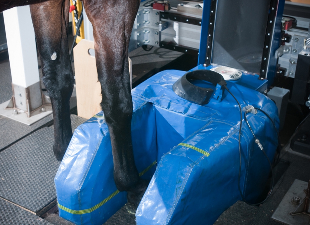
Standing MRI | UC Davis photo
Participants in the study-funded by the Oak Tree Charitable Foundation-will be split into two.
There will be 23 case horses whose lameness has been narrowed to the fetlock region, and 23 control horses who exhibit no visible sign of lameness.
The 23 case horses will be selected by Dr. Tim Grande, the California Horse Racing Board (CHRB)'s chief official veterinarian, from a variety of scenarios where regulatory veterinarians commonly have to intercede in a horse's training or racing program.
These include a morning or race-day scratch, a voided claim, and lameness in the test barn or following a scheduled work or race.
The control horses–those with no visible lameness–will be selected as a comparative match in terms of things like sex, age, and class.
If a case horse is picked from a race, then the winner–if sound–will make an obvious control match. If a case horse is selected after a workout, then a suitable match will be selected using PPs.
And what exactly will the researchers be looking for? The answer encompasses four specific areas of concern within the fetlock joint, the primary site of musculoskeletal injury in racehorses.
Researchers will be looking for density within the proximal sesamoid bones and distal cannon bone, bone marrow edema–or swelling–in the cannon bone, and palmer osteochondral disease, a type of bone bruising commonly referred to as just “POD.”
To elaborate on these points, Peloso pointed to a couple of relatively recent papers he had co-authored connecting important diagnostic dots.
Two issues associated with fetlock failure are high density–noticeable bone development that predisposes a horse to a greater risk of fracture–in the sesamoid bone and palmer osteochondral disease, while condylar fractures are linked to bone marrow edema and high density in the distal cannon bone.
“It's because of those two papers that we've dialed in on those bone changes,” said Peloso. “Maybe the study will teach us something new, and so, there'll be something that gets added to it.”
The standing MRI unit has been part of the Southern California backstretch furniture since the start of last year. Since then, the unit has been used to scan hundreds of fetlocks.
Nevertheless, as a relatively newfangled diagnostic tool, the MRI is still looked upon with a touch of skepticism by some corners of the backstretch community, including attending veterinarians, admitted Peloso.
As such, this study is seen as an opportunity to increase the volume of MRI equine traffic. “It needs to be a real relationship so we can do best by the horse and best by the owner,” Peloso explained, before looking at the broader implications from this and other such studies.
“It'll be interesting to see to what degree some of these imaging modalities–PET, CT if it makes it, MRI–what role they play to help the regulatory veterinarian identify who's safe and who's not safe.”
Not a subscriber? Click here to sign up for the daily PDF or alerts.
