
By Christina Bossinakis
Following the cancellation of the ninth Welfare and Safety of the June 20 Racehorse Summit in Lexington, Kentucky, because of the COVID-19 pandemic, the Grayson-Jockey Club Research Foundation kicked off an educational series of weekly webinars Tuesday afternoon. Presented by Dr. Katherine Garrett, shareholder at Rood & Riddle Equine Hospital, the initial segment on equine health and safety featured 'Fetlock Injuries: Palmar metacarpal disease and how the various imaging modalities can be used to help diagnose it.'
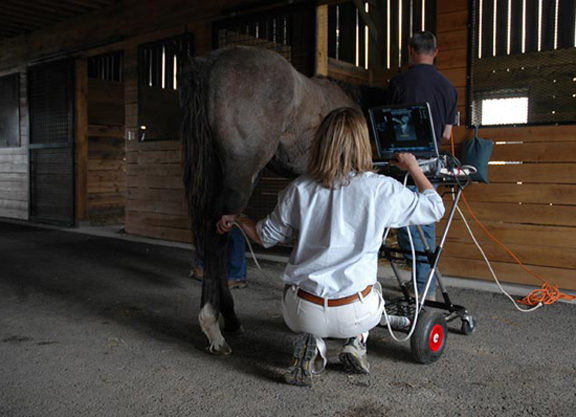
During the course of the 45-minute lecture, which was followed by a short Q&A period, Dr. Garrett outlined the different types of diagnostic imaging modalities–Radiography, Ultrasound, Nuclear Scintigraphy, MRI (Magnetic Resonance Imagining), CT (Computerized Tomography) and PET (Postitron Emission Tomography)–and the advantages and challenges inherent to each of them. The basic outline of each imaging modality and its best diagnostic use:
- Radiography – Good initial choice for bony problems
- Ultrasound – Good initial choice for soft tissue problems
- CT – Excellent choice for bony injury
- Nuclear Scintigraphy – (Functional imagining modality) Excellent choice for stress fractures, increased bony turnover
- MRI – Excellent choice for soft tissue and bony injury
- PET – (Functional imagining modality) – Excellent for evaluating bony turnover, higher resolution than scintigraphy and cross-sectional images (3D)
Dr. Garrett compared and contrasted each modality, when they are most effective, and how they are often used in tandem to provide the most accurate diagnosis.
“None of them can do everything,” said Garrett. “However, we look at Radiography and Ultrasonography and they shine in the convenience and expense categories. Most of the time, vets can make a diagnosis using Radiography or Ultrasonography, and that's why they're so common, because they are very good. But sometimes we do have to move to our more advanced imagining modalities. Looking at the modalities that have these advanced diagnostic abilities, often they cost more and the horse needs to go to a hospital or clinic where the service is provide. However, the trade-off for that is you're going to get the answer that you need, ideally.”
The webinar also offered a look at the general anatomy of the fetlock, in addition to a few of the injuries and diseases which affect the sport horse. Highlighted during the discussion were several commonly-seen injuries in racehorses, including Palmar Osteochondral Disease (POD), a condition that occurs due to the repetitive and high-speed concussion absorbed by the fetlock during training, resulting in the deterioration of the articular cartilage of the joint.
“POD develops when there is an imbalance between the work that is asked of the horse and the horse's ability to adapt the bone quickly enough,” explained Garrett. “Essentially, the bone can't keep up. And what this does is it leads to chronic subchondral bone fatigue, and the pain which manifests as lameness results from the damage to the subchondral bone.”
Addressing the challenge in diagnosing the condition, she continued, “There really is a spectrum of changes to the bone. This isn't normal one day and abnormal the next. This is part of a normal, adaptive training process, we expect to see some of these changes. However, the problem occurs when the bone isn't keeping up.”
Asked if imaging can predict lameness or injury, Dr. Garrett said, “Honestly, the answer right now is 'no'. The frustrating reason this is the case is because there is lot of overlap in the changes that are seen in a normal response to training and those seen in a pre-lameness or injury state. And there is a ton of variation between horses. That is why we need to treat the actual horse and not just the images.”
She added, “I suspect CT, MRI and PET scanning will play a major role in some of these endeavors. But currently, no test is 100% accurate. Several of the tests are very good, but asking for 100% accuracy is a lot. We really have to consider the horse as an individual.”
To watch Tuesday's webinar in its entirety, click here.
The next webinar in the series featuring–the Importance of transparency in medical records; monitoring horses between starts–will be held May 19. Moderating the segment is Dr. Dionne Benson, Chief Veterinary Officer, The Stronach Group and speakers include Dr. Ryan Carpenter, Equine Medical Center in Cypress, California; Dr. William Farmer, Equine Medical Director, Churchill Downs Inc.; and Dr. Scott Palmer, Equine Medical Director, New York State Gaming Commission.
The free webinars are accessible each week at zoom.us/j/96557992970. Viewers will be able to ask questions through the Zoom webinar platform. All sessions start at 2 p.m. ET.
Not a subscriber? Click here to sign up for the daily PDF or alerts.